

Accesory Structures Of The Skin
- Examine the anatomy of the integumentary system
- Decide the main functions of the integumentary system
- Differentiate integumentary system medical terms and common abbreviations
- Discover medical specialties associated with the integumentary system
- Recognize mutual diseases, disorders, and procedures related to the integumentary system
Integumentary Organisation Give-and-take Parts
Click on prefixes, combining forms, and suffixes to reveal a listing of discussion parts to memorize for the Integumentary Organisation.
Introduction to the Integumentary System
The integumentary arrangement refers to the skin and its accessory structures. In the adult homo body, the pare makes upwardly near 16% of body weight and covers an area of i.5 to 2 10002.
In fact, the pare and accessory structures are the largest organ system in the human body. The skin protects your inner organs, and information technology is in need of daily care and protection to maintain its health.
The skin and accessory structures are the largest organ system in the human being torso.
Watch this video:
Media five.1. The Integumentary Organization, Part ane – Skin Deep: Crash Form A&P #6 [Video]. Copyright 2015 by CrashCourse.
Exercise Medical Terms Related to the Integumentary System
Anatomy (Structures) of the Integumentary System
The skin and its accessory structures make up the integumentary system, which provides the body with overall protection. The peel is fabricated of multiple layers of cells and tissues, which are held to underlying structures by connective tissue (Effigy 5.one). The deeper layer of skin is well vascularized. It also has numerous sensory autonomic, and sympathetic nerve fibers ensuring communication to and from the brain.
The peel is composed of two primary layers:
- The epidermis
- The dermis
- Beneath the dermis lies the hypodermis, likewise known as the subcutaneous layer.
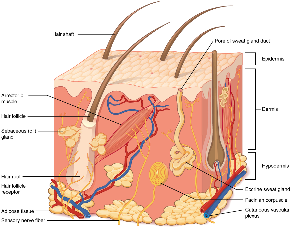
- On the diagram above discover the two layers of the skin: epidermis and dermis.
- The literal breakdown for hypodermis is "beneath the dermis." On the diagram to a higher place, where can you locate information technology?
- Tin can you find a pilus follicle , hair root, and hair shaft?
- Keep reading to find out what the arrector pili muscle does when you are frightened.
Epidermis
The epidermis is composed of keratinized, stratified squamous epithelium. It is made of four or five layers of epithelial cells, depending on its location in the body. It is avascular.
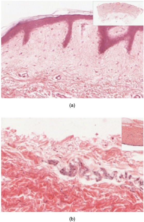
- Thin peel has four layers of cells. From deep to superficial, these layers are the stratum basale, stratum spinosum, stratum granulosum, and stratum corneum. Most of the skin can be classified as thin skin.
- Thick skin is found merely on the palms of the hands and the soles of the feet. It has a 5th layer, chosen the stratum lucidum, located betwixt the stratum corneum and the stratum granulosum (meet Figure 5.2).
The cells in all of the layers except the stratum basale are called keratinocytes. Keratin is an intracellular fibrous protein that gives hair, nails, and pare their hardness and water-resistant properties. The keratinocytes in the stratum corneum are expressionless and regularly slough abroad, being replaced by cells from the deeper layers (meet Figure 5.iii).
Dermis
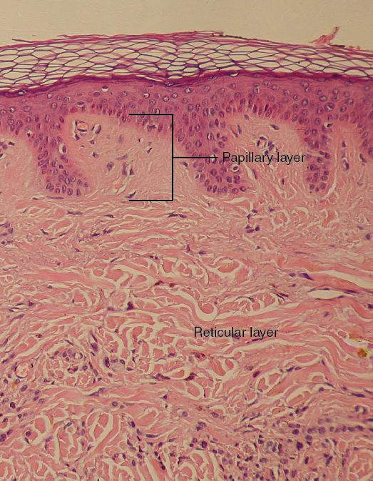
Papillary Layer
The papillary layer is fabricated of loose, areolar connective tissue, which means the collagen and elastin fibers of this layer course a loose mesh. This superficial layer of the dermis projects into the stratum basale of the epidermis to form finger-like dermal papillae (see Figure 5.four). Within the papillary layer are fibroblasts, a small number of adipocytes, and an abundance of small claret vessels. In improver, the papillary layer contains phagocytes that help fight bacteria or other infections that accept breached the skin. The layer also contains lymphatic capillaries, nerve fibers, and Meissner corpuscles.
Reticular Layer
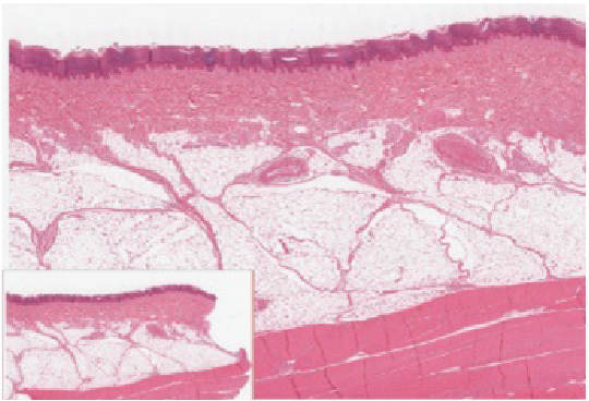
Underlying the papillary layer is the much thicker reticular layer, equanimous of dense, irregular connective tissue. The layer is well vascularized and has a rich sensory and sympathetic nervus supply. The reticular layer appears reticulated due to a tight meshwork of fibers. Elastin fibers provide some elasticity to the skin, enabling movement. Collagen fibers provide construction and tensile force, with strands of collagen extending into both the papillary layer and the hypodermis. In improver, collagen binds water to go on the skin hydrated. Collagen injections and Retin-A creams help restore skin turgor by either introducing collagen externally or stimulating blood flow and repair of the dermis, respectively.
Hypodermis (Subcutaneous Layer)
The hypodermis, also known every bit the subcutaneous layer, serves to connect the skin to the underlying fascia of the bones and muscles. Information technology is not strictly a function of the skin, although the border between the hypodermis and dermis can be difficult to distinguish. The hypodermis consists of well-vascularized, loose, areolar connective tissue and adipose tissue, which functions as a manner of fat storage and provides insulation and cushioning for the integument.
Practice Labeling the Layers of the Pare
Physiology (Role) of the Integumentary System
The skin and accessory structures perform a diverseness of essential functions, such equally protecting the body from invasion by microorganisms, chemicals, and other environmental factors; preventing aridity; acting equally a sensory organ; modulating body temperature and electrolyte balance; and synthesizing vitamin D. The underlying hypodermis has of import roles in storing fats, forming a "cushion" over underlying structures, and providing insulation from cold temperatures.
Protection
The skin protects the body from wind, h2o, and UV sunlight. It acts every bit a protective bulwark confronting water loss and information technology too is the beginning line of defense against abrasive activity such as dust, microbes, or harmful chemicals. Sweat excreted from sweat glands deters microbes from over-colonizing the skin surface past generating dermcidin, which has antibody properties.
Sensory Function
The skin acts as a sense organ because the epidermis, dermis, and hypodermis incorporate specialized sensory nerve structures that notice touch, surface temperature, and pain. These receptors are more concentrated on the tips of the fingers, which are virtually sensitive to impact, especially the Meissner corpuscle, which responds to lite touch, and the Pacinian corpuscle, which responds to vibration. Merkel cells, seen scattered in the stratum basale, are too touch receptors. In addition to these specialized receptors, in that location are sensory fretfulness continued to each hair follicle, hurting and temperature receptors scattered throughout the pare, and motor nerves innervate the arrector pili muscles and glands. This rich innervation helps u.s.a. sense our surroundings and react accordingly,
Thermoregulation
The integumentary arrangement helps regulate body temperature through its tight association with the sympathetic nervous organisation. The sympathetic nervous system is continuously monitoring trunk temperature and initiating appropriate motor responses.
- When the body becomes warm sweat glands, accompaniment structures to the peel, secrete water, salt, and other substances to absurd the trunk.
- Even when the body does not appear to exist noticeably sweating, approximately 500 mL of sweat are secreted a day.
- If the trunk becomes excessively warm due to high temperatures, vigorous activeness, or a combination of the two, sweat glands will be stimulated by the sympathetic nervous arrangement to produce large amounts of sweat.
- When the sweat evaporates from the skin surface, the body is cooled as body heat is prodigal.
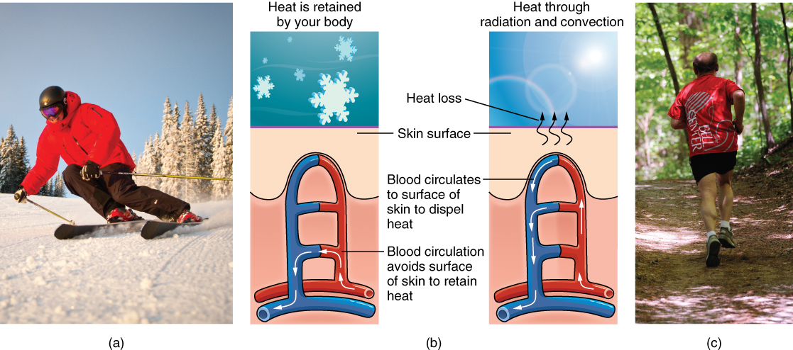
- In addition to sweating, arterioles in the dermis dilate and so that excess heat carried by the blood tin can dissipate through the skin and into the surrounding environment (Figure 5.5).
- This accounts for the skin redness that many people experience when exercising.
- When body temperatures driblet, the arterioles constrict to minimize heat loss, peculiarly in the ends of the digits and tip of the nose.
- This reduced circulation tin effect in the skin taking on a whitish hue.
- Although the temperature of the peel drops as a result, passive heat loss is prevented, and internal organs and structures remain warm.
- If the temperature of the pare drops besides much (such equally environmental temperatures below freezing), the conservation of body core oestrus can issue in frostbite.
Can you describe the thermoregulation process between the integumentary system and the sympathetic arrangement?
- What happens when the body temperature is too warm?
- What happens when the trunk temperature is too cold?
Vitamin D Synthesis
- Vitamin D is essential for the normal absorption of calcium and phosphorus, which are required for salubrious bones.
- The absence of dominicus exposure can lead to a lack of vitamin D in the body. In children, this can cause rickets. Vitamin D deficiency in elderly individuals may pb to osteomalacia.
- In present-day social club, Vitamin D is added equally a supplement to many foods, including milk and orange juice, compensating for the need for sun exposure. In addition to its essential role in bone health, Vitamin D is essential for full general immunity against bacterial, viral, and fungal infections.
Vitamin D is essential for general immunity against bacterial, viral, and fungal infections.
Sentinel this video:
Media 5.2. The Integumentary System, Function 2 – Skin Deeper: Crash Course A&P #vii [Online video]. Copyright 2015 by CrashCourse.
Accessory Structures
Accessory structures of the pare include hair, nails, sweat glands, and sebaceous glands. These structures embryologically originate from the epidermis and can extend downwards through the dermis into the hypodermis.
Hair
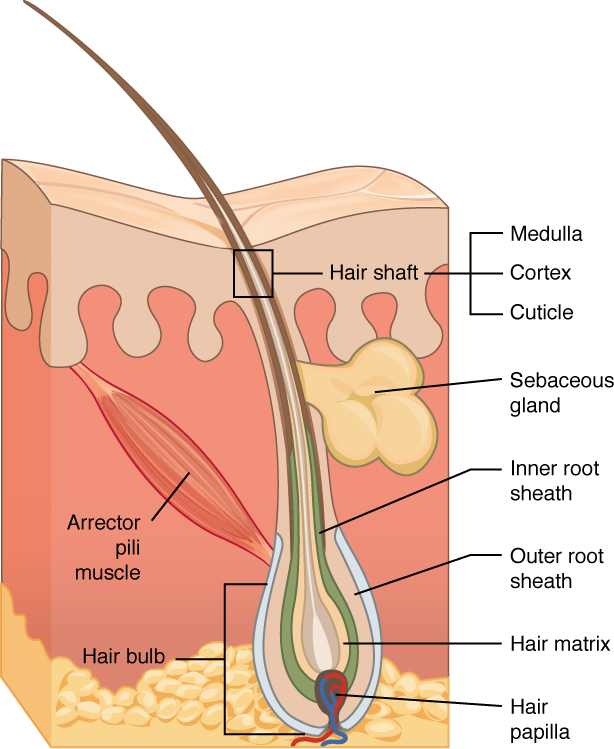
Hair is a keratinous filament growing out of the epidermis. Information technology is primarily fabricated of dead, keratinized cells. Strands of hair originate in an epidermal penetration of the dermis called the hair follicle. The pilus shaft is the function of the hair non anchored to the follicle, and much of this is exposed at the skin'south surface. The residuum of the hair, which is anchored in the follicle, lies below the surface of the skin and is referred to as the hair root. The pilus root ends deep in the dermis at the pilus bulb and includes a layer of mitotically agile basal cells called the hair matrix. The hair bulb surrounds the hair papilla, which is made of connective tissue and contains claret capillaries and nerve endings from the dermis (see Effigy 5.half-dozen).
Pilus Function
Hair serves a variety of functions, including protection, sensory input, thermoregulation, and communication. For example:
- Hair on the head protects the skull from the dominicus.
- Hair in the nose and ears, and around the eyes (eyelashes) defends the body past trapping and excluding dust particles that may contain allergens and microbes.
- Hair of the eyebrows prevents sweat and other particles from dripping into and bothering the eyes.
Pilus also has a sensory function due to sensory innervation by a hair root plexus surrounding the base of each hair follicle. Hair is extremely sensitive to air movement or other disturbances in the surround, much more and then than the skin surface. This feature is also useful for the detection of the presence of insects or other potentially damaging substances on the skin surface.
Each hair root is connected to a smooth musculus called the arrector pili that contracts in response to nervus signals from the sympathetic nervous system, making the external pilus shaft "stand up up." The master purpose for this is to trap a layer of air to add insulation. This is visible in humans as goosebumps. Of course, this is much more obvious in organisms with a heavier coat than most humans, such as when a frightened cat raises its fur.
When frightened, the arrector pili muscle is responsible for your pilus standing on terminate. The same is true when a true cat's fur is raised.
Hair Growth, Loss, and Colour
Hair grows and is eventually shed and replaced by new hair. Hair typically grows at the charge per unit of 0.three mm per day. On average, l hairs are lost and replaced per twenty-four hour period. Hair loss occurs if there is more than hair shed than what is replaced and can happen due to hormonal or dietary changes. Hair loss tin can besides result from the crumbling process, or the influence of hormones. Similar to the skin, pilus gets its color from the paint melanin, produced by melanocytes in the hair papilla. Different hair colour results from differences in the blazon of melanin. Equally a person ages, the melanin product decreases, and pilus tends to lose its color and becomes gray and/or white.
Nails
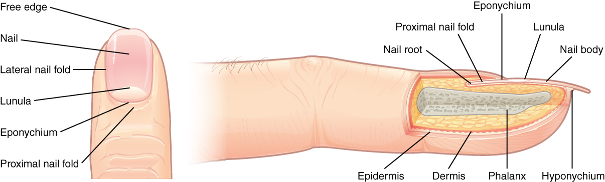
The nail bed is a specialized construction of the epidermis that is institute at the tips of our fingers and toes. The blast trunk is formed on the nail bed and protects the tips of our fingers and toes every bit they are the uttermost extremities and the parts of the torso that feel the maximum mechanical stress (see Figure 5.seven). The boom body forms a back-support for picking upwards small objects with the fingers. The boom body is composed of densely packed dead keratinocytes.
The epidermis in this office of the body has evolved a specialized structure upon which nails can course. The smash body forms at the nail root, which has a matrix of proliferating cells from the stratum basale that enables the nail to abound continuously. The lateral nail fold overlaps the nail on the sides, helping to anchor the nail trunk. The nail fold that meets the proximal terminate of the boom body forms the nail cuticle, also called the eponychium.
The blast bed is rich in blood vessels, making it announced pink, except at the base where a thick layer of epithelium over the nail matrix forms a crescent-shaped region chosen the lunula (the "little moon"). The surface area beneath the free border of the nail, furthest from the cuticle, is chosen the hyponychium. It consists of a thickened layer of stratum corneum.
Sweat Glands
Sudoriferous Glands
When the body becomes warm, sudoriferous glands produce sweat to absurd the trunk. Sweat glands develop from epidermal projections into the dermis and are classified every bit merocrine glands; that is, the secretions are secreted by exocytosis through a duct without affecting the cells of the gland. There are two types of sweat glands, each secreting slightly unlike products.
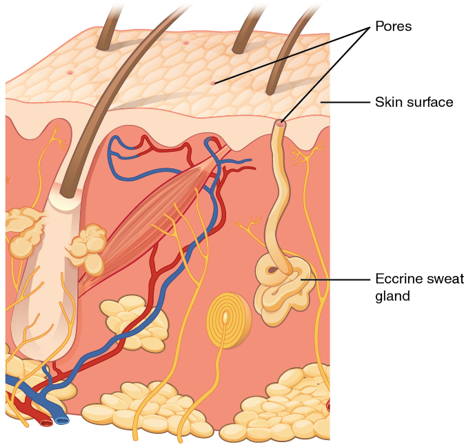
An eccrine sweat gland is a type of gland that produces hypotonic sweat for thermoregulation as described previously. These glands are institute all over the pare'south surface just are especially arable on the palms of the hand, the soles of the feet, and the forehead (Figure five.8). They are coiled glands lying deep in the dermis, with the duct ascent upwardly to a pore on the skin surface where the sweat is released. This type of sweat, released by exocytosis, is hypotonic and equanimous mostly of water, with some salt, antibodies, traces of metabolic waste, and dermcidin, an antimicrobial peptide. Eccrine glands are a main component of thermoregulation in humans and thus assistance to maintain homeostasis.
An apocrine sweat gland is normally associated with hair follicles in densely hairy areas, such as armpits and genital regions. Apocrine sweat glands are larger than eccrine sweat glands and prevarication deeper in the dermis, sometimes even reaching the hypodermis, with the duct usually elimination into the pilus follicle. In addition to water and salts, apocrine sweat includes organic compounds that make the sweat thicker and subject to bacterial decomposition and subsequent smell. The release of this sweat is under both nervous and hormonal control and plays a function in the poorly understood human pheromone response. Most commercial antiperspirants use an aluminum-based compound as their primary active ingredient to stop sweat. When the antiperspirant enters the sweat gland duct, the aluminum-based compounds precipitate due to a change in pH and course a physical block in the duct, which prevents sweat from coming out of the pore.
Sebaceous Glands
A sebaceous gland is a type of oil gland that is found all over the body and helps to lubricate and waterproof the skin and pilus. About sebaceous glands are associated with pilus follicles. They generate and excrete sebum, a mixture of lipids, onto the skin surface, thereby naturally lubricating the dry and dead layer of keratinized cells of the stratum corneum, keeping it pliable. The fatty acids of sebum also take antibacterial properties and forestall water loss from the skin in low-humidity environments. The secretion of sebum is stimulated past hormones, many of which do not go active until puberty. Thus, sebaceous glands are relatively inactive during childhood.
Aluminum-based compounds due to a change in pH form a physical block in the sweat gland duct. This prevents sweating.
Practice Terms Related to the Integumentary System
Common Abbreviations for the Integumentary Organisation
Many terms and phrases related to the integumentary system are abbreviated. Larn these common abbreviations by expanding the list below.
Changes Due to Aging
All systems in the trunk accumulate subtle and some not-so-subtle changes as a person ages. Among these changes are reductions in jail cell division, metabolic activity, blood apportionment, hormonal levels, and muscle forcefulness (encounter Figure 5.9). In the skin, these changes are reflected in decreased mitosis in the stratum basale, leading to a thinner epidermis. The dermis, which is responsible for the elasticity and resilience of the peel, exhibits a reduced ability to regenerate, which leads to slower wound healing. The hypodermis, with its fatty stores, loses construction due to the reduction and redistribution of fat, which in plow contributes to the thinning and sagging of peel.
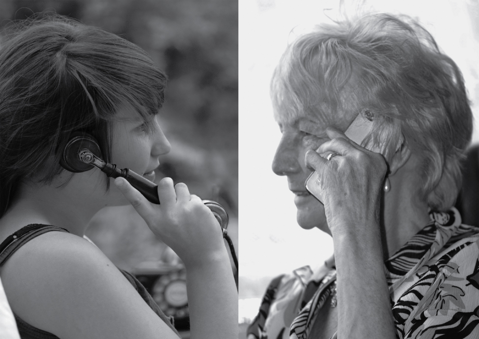
The accompaniment structures also accept lowered activity, generating thinner pilus and nails, and reduced amounts of sebum and sweat. A reduced sweating ability can cause some elderly to be intolerant to extreme heat. Other cells in the skin, such as melanocytes and dendritic cells, likewise become less active, leading to a paler pare tone and lowered amnesty. Wrinkling of the skin occurs due to the breakdown of its construction, which results from decreased collagen and elastin production in the dermis, weakening of muscles lying under the skin, and the disability of the skin to retain adequate moisture.
A reduced sweating power tin crusade some older adults to be intolerant to extreme heat.
Diseases and Disorders of the Integumentary System
The integumentary organization is susceptible to a diversity of diseases, disorders, and injuries. These range from annoying but relatively beneficial bacterial or fungal infections that are categorized equally disorders, to skin cancer and severe burns, which can exist fatal. In this section, you will learn several of the most common skin atmospheric condition.
Most cancers are identified by the organ or tissue in which the cancer originates. In general, cancers effect from an accumulation of Dna mutations. These mutations can result in cell populations that do non die when they should and uncontrolled cell proliferation that leads to tumors. Although many tumors are benign, some metastasize. Cancers are characterized by their power to metastasize. One common class of cancer is skin cancer.
Sun Harm
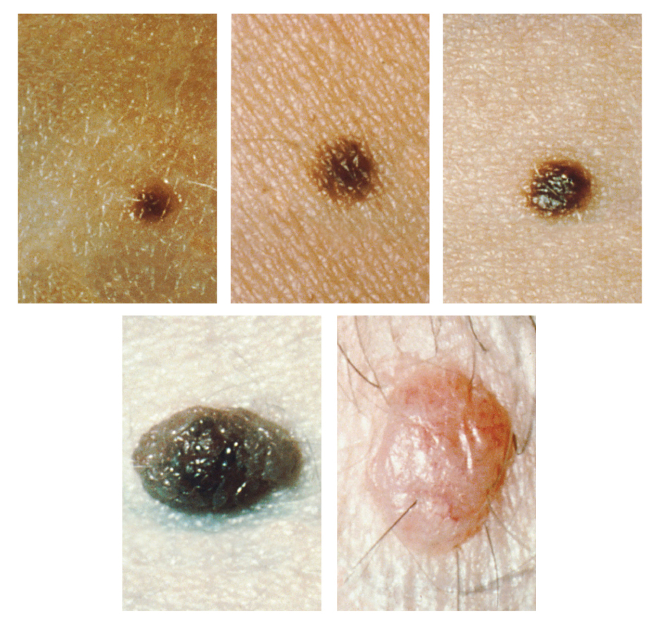
Melanin synthesis peaks well-nigh 10 days after initial sun exposure, which is why pale-skinned individuals tend to endure sunburns of the epidermis initially. Dark-skinned individuals can also get sunburns but are more protected than are pale-skinned individuals. Too much sun exposure tin can eventually atomic number 82 to wrinkling due to the devastation of the cellular construction of the peel, and in severe cases, tin cause sufficient Deoxyribonucleic acid impairment to result in skin cancer. When there is an irregular aggregating of melanocytes in the pare, freckles appear. Moles are larger masses of melanocytes, and although almost are beneficial, they should be monitored for changes that might indicate the presence of cancer (see Figure 5.10).
Basal Cell Carcinoma (BCC)
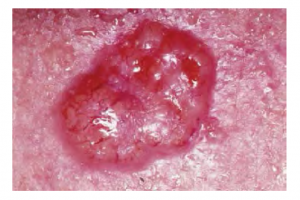
Basal jail cell carcinoma is a form of cancer that affects the mitotically active stem cells in the stratum basale of the epidermis. It is the near mutual of all cancers that occur in the United States and is frequently establish on the head, neck, arms, and back, which are the well-nigh susceptible to long-term sun exposure. Although UV rays are the main culprit, exposure to other agents, such equally radiations and arsenic, can also lead to this blazon of cancer. Wounds on the skin due to open sores, tattoos, burns, et cetera may be predisposing factors. Basal cell carcinomas starting time in the stratum basale and usually spread along this boundary. At some point, they begin to grow toward the surface and go an uneven patch, bump, growth, or scar on the pare surface (see Figure five.eleven). Like most cancers, basal cell carcinomas reply best to treatment when caught early. Treatment options include surgery, freezing (cryosurgery), and topical ointments.
Squamous Cell Carcinoma (SCC)
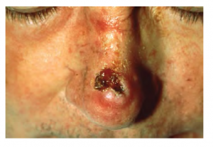
Squamous prison cell carcinoma is cancer that affects the keratinocytes of the stratum spinosum and presents equally lesions commonly found on the scalp, ears, and easily (see Figure 5.12). Information technology is the 2nd most common skin cancer. The American Cancer Society reports that two of 10 skin cancers are squamous cell carcinomas, and information technology is more than aggressive than basal cell carcinoma. If not removed, these carcinomas can metastasize. Surgery and radiation are used to cure squamous cell carcinoma.
Melanoma
Melanoma is cancer characterized by the uncontrolled growth of melanocytes, the pigment-producing cells in the epidermis. Typically, a melanoma develops from a mole. Information technology is the most fatal of all peel cancers, every bit information technology is highly metastatic and can be hard to detect before it has spread to other organs. Melanomas unremarkably announced as asymmetrical brown and black patches with uneven borders and a raised surface (see Figure v.13). Treatment typically involves surgical excision and immunotherapy.
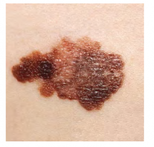
ABCDE for Early Diagnosis
Doctors oftentimes give their patients the following ABCDE mnemonic to assistance with the diagnosis of early on-stage melanoma. If you observe a mole on your body displaying these signs, consult a md:
- Asymmetry: the two sides are not symmetrical
- Borders: the edges are irregular in shape
- Color: the colour is varied shades of brown or black
- Diameter: information technology is larger than six mm (0.24 in)
- Eastvolving: its shape has changed
Some specialists cite the post-obit boosted signs for the most serious course, nodular melanoma:
- Elevated: information technology is raised on the skin surface
- Firm: it feels hard to the bear on
- Growing: it is getting larger
Albinism
Albinism is a genetic disorder that affects (completely or partially) the coloring of skin, hair, and eyes. This is primarily due to the inability of melanocytes to produce melanin. Individuals with albinism tend to appear white or very pale due to the lack of melanin in their skin and pilus. Call up that melanin helps protect the skin from the harmful furnishings of UV radiation. Individuals with albinism tend to need more than protection from UV radiation, as they are more prone to sunburns and skin cancer. They also tend to exist more than sensitive to light and have vision bug due to the lack of pigmentation on the retinal wall.
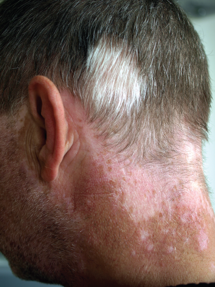
Treatment of this disorder usually involves addressing the symptoms, such as limiting UV low-cal exposure to the skin and eyes. In vitiligo , the melanocytes in certain areas lose their ability to produce melanin, possibly due to an autoimmune reaction. This leads to a loss of color in patches (see Figure 5.14). Neither albinism nor vitiligo directly affects the lifespan of an individual.
Changes in Skin Coloration
Other changes in the advent of skin coloration can be indicative of diseases associated with other body systems.
- Liver disease or liver cancer can crusade the accumulation of bile and the yellow pigment bilirubin, leading to the skin appearing yellow or jaundiced.
- Tumors of the pituitary gland can issue in the secretion of large amounts of melanocyte-stimulating hormone (MSH), which results in a concealment of the skin.
- Addison's illness can stimulate the release of backlog amounts of adrenocorticotropic hormone (ACTH), which tin can requite the peel a deep statuary colour
- A sudden driblet in oxygenation can touch pare color, causing the peel to initially turn cadaverous (white).
- Subsequently a prolonged reduction in oxygen levels, dark ruby-red deoxyhemoglobin becomes dominant in the blood, making the skin appear blue, a condition referred to every bit cyanosis. This happens when the oxygen supply is restricted, as when someone is experiencing difficulty in breathing because of asthma or a center attack. Withal, in these cases, the outcome on skin color has nothing to do with the pare's pigmentation.
Skin Disorders
Ii common peel disorders are eczema and acne. Eczema is an inflammatory status and occurs in individuals of all ages. Acne involves the bottleneck of pores, which can lead to infection and inflammation and is ofttimes seen in adolescents. Other disorders include seborrheic dermatitis (on the scalp), psoriasis, fungal infections, cold sores, impetigo, scabies, hives, and warts.
Eczema
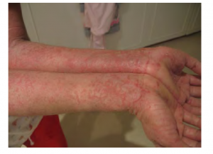
Eczema is an allergic reaction that manifests every bit dry, itchy patches of skin that resemble rashes (see Effigy five.15). It may be accompanied past swelling of the skin, flaking, and in severe cases, bleeding. Symptoms are usually managed with moisturizers, corticosteroid creams, and immunosuppressants.
Acne
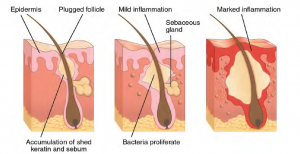
Acne is a peel disturbance that typically occurs on areas of the skin that are rich in sebaceous glands (face and dorsum). It is nigh mutual along with the onset of puberty due to associated hormonal changes, simply can as well occur in infants and continue into adulthood. Hormones, such as androgens, stimulate the release of sebum. Overproduction and accumulation of sebum along with keratin tin block pilus follicles. This plug is initially white. The sebum, when oxidized past exposure to air, turns black. Acne results from infection past acne-causing bacteria (Propionibacterium and Staphylococcus), which tin lead to redness and potential scarring due to the natural wound healing process (see Figure v.16).
Ringworm
Tinea or dermatophytosis is often referred to as ringworm. Ringworm presents every bit a circular rash that is itchy and ruby-red and can be found on diverse parts of the body. It is referred to by the location that it is found:
- Tinea pedis – feet (usually referred to every bit athlete's anxiety)
- Tinea capitis – scalp
- Tinea barbae – bristles
- Tinea manuum – hands
- Tinea unguium – toenails and fingernails (also chosen onychomycosis)
- Tinea corporis – body parts such every bit artillery and legs (Centre for Disease Control and Prevention, n.d.-a)
To learn more virtually ringworm, visit the Center for Disease Control and Prevention's web folio on fungal infections.
Psoriasis
Psoriasis is a chronic autoimmune disorder that results in patches of thick red pare with the advent of silverish scales. These patches tin can be institute on elbows, knees, scalp, low back, face, feet, fingernails, toenails, and even the mouth. Psoriasis can be confused with other peel diseases, and then a dermatologist is the best doc to diagnose psoriasis. Treatments may include creams, ointments, ultraviolet light therapy, and medication (Center for Affliction Control and Prevention, n.d.-b). To learn more, visit the Center for Disease Control and Prevention'south web folio on psoriasis.
Injuries
Skin injuries prepare off a healing process that occurs in several overlapping stages.
- The kickoff footstep to repairing damaged skin is the formation of a blood jell that helps terminate the menstruum of claret and scabs over time. Many dissimilar types of cells are involved in wound repair, especially if the expanse that needs repair is extensive.
- Earlier the basal stalk cells of the stratum basale tin can recreate the epidermis, fibroblasts mobilize and divide chop-chop to repair the damaged tissue by collagen deposition, forming granulation tissue.
- Blood capillaries follow the fibroblasts and help increment blood circulation and oxygen supply to the area.
- Immune cells, such equally macrophages, roam the area and engulf any strange matter to reduce the run a risk of infection.
Burns
A burn results when the skin is damaged by intense rut, radiations, electricity, or chemicals. The damage results in the death of peel cells, which can lead to a massive loss of fluid. Dehydration, electrolyte imbalance, and renal and circulatory failure follow, which can exist fatal. Fire patients are treated with intravenous fluids to showtime aridity, as well as intravenous nutrients that enable the body to repair tissues and supersede lost proteins. Another serious threat to the lives of burn down patients is infection. Burned skin is extremely susceptible to leaner and other pathogens due to the loss of protection by intact layers of skin.
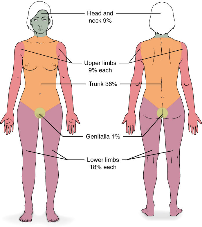
Burn Classification
Burns are sometimes measured in terms of the size of the total surface surface area affected. This is referred to as the rule of nines, which associates specific anatomical areas with a percentage that is a cistron of nine (see Figure v.17).
Burns are also classified by the degree of their severity:
- A first-caste burn is a superficial burn that affects only the epidermis. Although the skin may be painful and swollen, these burns typically heal on their own within a few days. Mild sunburn fits into the category of a offset-degree fire.
- A 2d-degree burn goes deeper and affects both the epidermis and a portion of the dermis. These burns upshot in swelling and a painful baking of the skin. It is of import to go on the burn down site clean and sterile to prevent infection. If this is done, the burn will heal within several weeks.
- A third-degree burn fully extends into the epidermis and dermis, destroying the tissue and affecting the nerve endings and sensory part. These are serious burns that may appear white, red, or black; they require medical attention and will heal slowly without information technology.
- A fourth-caste burn is even more than severe, affecting the underlying muscle and bone.
Oddly, 3rd and fourth-degree burns are usually not as painful considering the nerve endings themselves are damaged. Total-thickness burns cannot be repaired by the body, considering the local tissues used for repair are damaged and require debridement, or amputation in severe cases, followed by grafting of the skin from an unaffected part of the torso, or from skin grown in tissue culture for grafting purposes. Skin grafts are required when the damage from trauma or infection cannot exist airtight with sutures or staples.
Scars and Keloids
Most cuts or wounds, with the exception of ones that simply scratch the epidermis, atomic number 82 to scar germination. Scarring occurs in cases in which at that place is repair of skin damage, but the skin fails to regenerate the original skin structure. Fibroblasts generate scar tissue in the form of collagen, and the bulk of repair is due to the basket-weave pattern generated past collagen fibers and does not issue in regeneration of the typical cellular structure of skin. Instead, the tissue is fibrous in nature and does non let for the regeneration of accessory structures, such as pilus follicles, sweat glands, or sebaceous glands.
Sometimes, at that place is an overproduction of scar tissue, because the process of collagen germination does not stop when the wound is healed; this results in a keloid. In contrast, scars that issue from acne and chickenpox have a sunken appearance and are called atrophic scars.
Scarring of pare after wound healing is a natural process and does not demand to be treated farther. Application of mineral oil and lotions may reduce the formation of scar tissue. However, modern cosmetic procedures, such as dermabrasion, light amplification by stimulated emission of radiation treatments, and filler injections have been invented every bit remedies for severe scarring. All of these procedures try to reorganize the structure of the epidermis and underlying collagen tissue to make information technology wait more than natural.
Bedsores and Stretch Marks
Pare and its underlying tissue tin can be affected past excessive pressure. One example of this is called a bedsore. Bedsores, besides called decubitus ulcers, are caused by abiding, long-term, unrelieved pressure on certain body parts that are bony, reducing claret catamenia to the area and leading to necrosis. Bedsores are nigh common in elderly patients who have debilitating conditions that crusade them to be immobile. Near hospitals and long-term intendance facilities have the do of turning the patients every few hours to prevent the incidence of bedsores. If left untreated, bedsores tin be fatal if they go infected.
The peel can likewise be afflicted by pressure associated with rapid growth. A stretch mark results when the dermis is stretched across its limits of elasticity, every bit the skin stretches to arrange the backlog pressure. Stretch marks usually accompany rapid weight gain during puberty and pregnancy. They initially have a reddish hue but lighten over time. Other than for corrective reasons, treatment of stretch marks is not required. They occur most unremarkably over the hips and abdomen.
Calluses
When you wear shoes that do not fit well and are a abiding source of abrasion on your toes, y'all tend to grade a callus at the point of contact. This occurs because the basal stem cells in the stratum basale are triggered to divide more often to increment the thickness of the peel at the point of chafe to protect the residuum of the trunk from farther harm. This is an example of a pocket-sized or local injury, and the peel manages to react and treat the problem independently of the residuum of the body. Calluses can also form on your fingers if they are bailiwick to constant mechanical stress, such as long periods of writing, playing string instruments, or video games. A corn is a specialized form of callus. Corns form from abrasions on the skin that result from an elliptical-type motion.
Medical Terms in Context
Medical Specialties and Procedures Related to the Integumentary System
A dermatologist is a medical physician with specialized grooming in treating diseases, disorders, and injuries related to the integumentary system and its accessory structures. There are many dermatologic subspecialties, such as corrective dermatology, dermatopathology, and pediatric dermatology. To learn more, visit the American Academy of Dermatology Association's webpage on dermatology as a career.
Dermatologists can exist specially trained to perform a procedure called Mohs surgery. Mohs surgery excises peel cancers in thin layers until all cancer is removed from the tissue (Prickett & Ramsey, 2021).
Integumentary System Vocabulary
Abscess
An enclosed collection of pus in tissues, organs, or bars spaces in the torso.
Adipocyte
Fat jail cell.
Adipose tissue
Fat tissue.
Autonomic
Involuntary or unconscious.
Avascular
Without blood vessels.
Bacteria
Single-cell microorganisms that reproduce by cell sectionalisation and may cause infection by invading trunk tissue.
Benign
Non-cancerous.
Biopsy
The removal of cells or tissues for examination by a pathologist.
Cancer
Abnormal cells in the body that divide uncontrollably.
Cauterize
To destroy tissue using a hot or cold instrument, an electrical current, or a chemical that burns or dissolves the tissue to kill tumors or cease haemorrhage.
Cellulitis
An infection of the skin and subcutaneous tissue, characterized past tenderness, fever, and blisters.
Contusion
Injury resulting in a bruise.
Cyanosis
A condition in which the oxygen supply is restricted, causing the pare to look blue.
Cyst
Closed sac containing fluid or semisolid material.
Debridement
Excision of damaged tissues and cell debris from a wound or fire to foreclose infection and promote healing.
Dehydration
A net loss of water that results in insufficient water in blood and other tissues.
Dermabrasion
A procedure to remove superficial scars using sandpaper or revolving wire brushes.
Dermatitis
Inflammation of the pare.
Dermatofibroma
Gristly tumor of the peel.
Dermatologist
Medical doctor who specializes in diagnosing and treating skin disorders.
Dermatology
Study of disorders of the skin.
Dermis
The layer of skin that is made of dense, irregular connective tissue that houses claret vessels, hair follicles, sweat glands, and other structures.
Diaphoresis
Sweating.
Eczema
Non-infectious, inflammatory disease presenting as redness, blisters, scabs, and itching.
Edema
Swelling due to excessive liquid in the tissues.
Epidermis
The outer, protective layer of the skin.
Excisional skin surgery
A surgical procedure used to remove moles, cysts, skin cancer, and other pare growths using local anesthesia.
Exocytosis
A grade of active transport in which a cell exports textile using vesicular send.
Fascia
Fibrous tissue.
Frostbite
A condition in which conservation of the body cadre heat results in the skin freezing.
Gangrene
Expiry of tissue due to claret supply loss.
Hidradenitis
Inflammation of a sweat gland.
Hypodermis
Also known every bit the subcutaneous layer; the layer of the skin below the dermis that is composed mainly of loose connective and fatty tissues.
Incision
A cut made in the body to perform surgery.
Infection
The invasion and growth of bacteria, viruses, yeast, fungi, or other microorganisms in the body.
Intradermal
Within the skin.
Intravenous
Into or within the vein.
Jaundiced
Yellowish-colored.
Keloid
A raised or hypertrophic scar.
Keratinocytes
Cells that industry and shop the protein keratin.
Keratosis
Any growth of horny tissue.
Laceration
Torn, ragged-edged wound.
Lesion
An area of abnormal tissue.
Meissner corpuscle
A specialized sensory nerve structure that responds to lite touch.
Melanocyte
A cell that produces the pigment melanin.
Metastasis
The process in which cancer spreads from i office of the torso to another.
Necrosis
Accidental cell expiry.
Nevus
A benign growth on the skin that is formed by a cluster of melanocytes.
Nodule
A growth or lump that may be malignant or benign.
Onychocryptosis
An ingrown boom.
Onychomycosis
A fungal infection of the blast.
Onychophagia
Blast-bitter.
Osteomalacia
A softening of adult bones due to Vitamin D deficiency.
Pacinian corpuscle
A specialized sensory nerve structure that responds to vibration.
Pallor
Unnatural paleness of the skin.
Paronychia
Infection of the pare effectually the nail.
Pathogen
An organism that causes a disease.
Percutaneous
Passing through the skin, every bit an injection or a topical medicine.
Phagocytes
Cells that engulf and absorb bacteria and jail cell particles.
Pruritus
Itching.
Reticulated
Internet like.
Rhytidoplasty
Excision of wrinkles of the peel.
Rickets
A painful condition in children where bones are misshapen due to a lack of calcium, causing bow-leggedness.
Scar
A collagen-rich skin formed after the procedure of wound healing that differs from normal skin.
Staphylococcus aureus
A bacteria that is usually found in minor peel infections, also as in the nose of some good for you people.
Stratum basale
The deepest layer of the epidermis.
Streptococcus
The leaner that causes strep throat.
Subcutaneous
Below the skin.
Sympathetic nervous system
The division of the nervous system involved in our fight-or-flight responses. It continuously monitors body temperature and initiates appropriate motor responses.
Tinea
A grouping of fungal peel diseases of the hair, peel, and nail tissues.
Transdermal
Captivated through the unbroken skin.
Vascularized
Tissue that has numerous blood vessels.
Virus
A simple microorganism that may crusade infection by invading trunk tissue.
Exam Yourself
References
Centers for Illness Control and Prevention. (n.d.-a). Ringworm. Centers for Disease Control and Prevention: Fungal Diseases. https://www.cdc.gov/fungal/diseases/ringworm/definition.html
Centers for Disease Control and Prevention. (northward.d.-b). Psoriasis. Centers for Disease Control and Prevention: Fungal Diseases. https://world wide web.cdc.gov/psoriasis/
CrashCourse. (2015, January vi). The Integumentary system, part ane – peel deep: Crash Course A&P #6 [Video]. YouTube. https://youtu.be/Orumw-PyNjw
CrashCourse. (2015, Feb xvi). The Integumentary organization, role 2 – pare deeper: Crash Course A&P #7 [Video]. YouTube. https://youtu.be/EN-10-zXXVwQ
Prickett, K. A., & Ramsey, M. L. (2021). Mohs micrographic surgery. In StatPearls [Internet]. https://www.ncbi.nlm.nih.gov/books/NBK441833/
Image Descriptions
Figure 5.1 prototype clarification: This analogy shows a cross-section of pare tissue. The outermost layer is chosen the epidermis and occupies one-fifth of the cantankerous-section. Several hairs are emerging from the surface. The epidermis dives around one of the hairs, forming a follicle. The heart layer is chosen the dermis, which occupies 4-fifths of the cantankerous-department. The dermis contains an arrector pili muscle connected to one of the follicles. The dermis also contains an eccrine sweat gland, equanimous of a bunch of tubules. I tubule travels upward from the bunch, through the epidermis, opening onto the surface of a pore. There are 2 string-like nerves traveling vertically through the dermis. The right nerve is attached to a Pacinian corpuscle, which is a yellowish structure consisting of concentric ovals similar to an onion. The lowest level of the skin, the hypodermis, contains fatty tissue, arteries, and veins. Claret vessels travel from the hypodermis and connect to hair follicles and arrector pili muscle in the dermis. [Render to Figure 5.one].
Figure v.2 paradigm description: Role A is a micrograph showing a cantankerous-section of sparse skin. The topmost layer is a thin, translucent layer with irregular texture and areas where cells are sloughing off. The deepest layer is nighttime majestic and extends into the 3rd layer with finger-like projections. The 3rd lite purple layer contains sparse bands of fibers and small, dark cells. The fourth, and deepest layer, is darker than the tertiary layer merely is yet light regal. It contains thick fiber bands that are loosely packed. Part B is a magnified view of the epidermis of thick pare. Information technology shows the topmost layer is v times thicker than the topmost layer of sparse skin. The topmost layer of thick skin is likewise denser and less translucent than the topmost layer of thin skin. [Return to Effigy 5.2].
Figure 5.3 image description: The outer layer of cells in this micrograph is the thinnest layer and stained deep purple due to the full keratinization of expressionless cells. The adjacent layer occupies i-quarter of the micrograph, is lightly stained, and is a dense collection of cells. The third layer from the top is mostly white, with lightly stained, loosely-packed strands radiating in random directions. The bottom-most layer is densely-packed, with thick bands of highly organized muscle tissue that are darkly stained. [Return to Figure 5.3].
Figure five.four paradigm clarification: This micrograph shows layers of skin in a cross-department. The papillary layer of the dermis extends between the downward fingers of the darkly stained epidermis. The papillary layer appears effectively than the reticular layer, consisting of smaller, densely-packed fibers. The reticular layer is three times thicker than the papillary layer and contains larger, thicker fibers. The fibers seem more loosely packed than those of the papillary layer, with some separated past empty spaces. Both layers of the dermis contain cells with darkly stained nuclei. [Return to Effigy 5.iv].
Figure five.v epitome description: Part A is a photo of a homo skiing with several snow-covered trees in the background. Part B is a diagram with a correct and left one-half. The left half is titled " Heat is retained by the torso," while the right half is titled "Heat loss through radiations and convection." Both evidence blood flowing from an artery through iii capillary beds within the pare. The beds are arranged vertically, with the topmost bed located along the boundary of the dermis and epidermis. The bottommost bed is located deep in the hypodermis. The eye bed is evenly spaced between the topmost and bottommost beds. In each bed, oxygenated blood (red) enters the bed on the left and deoxygenated blood (blue) leaves the bed on the right. The left diagram shows a pic of snowflakes above the capillary beds, indicating that the weather is cold. Blood is only flowing through the deepest of the iii capillary beds, as the upper beds are closed off to reduce estrus loss from the outer layers of the skin. The right diagram shows a picture of the sunday above the capillary beds, indicating that the weather is hot. Blood is flowing through all three capillary beds, allowing heat to radiate out of the blood, increasing estrus loss. Part C is a photo of a man running through a forested trail on a summer day. [Return to Figure five.5].
Effigy five.half-dozen image description: A cross-section of the pare containing a hair follicle. The follicle is teardrop-shaped. Its enlarged base, labeled the hair bulb, is embedded in the hypodermis. The outermost layer of the follicle is the epidermis, which invaginates from the pare surface to envelop the follicle. Within the epidermis is the outer root sheath, which is simply present on the hair bulb. It does not extend up the shaft of the hair. Inside the outer root sheath is the inner root sheath. The inner root sheath extends nigh one-half of the way up the hair shaft, ending midway through the dermis. The pilus matrix is the innermost layer. The hair matrix surrounds the bottom of the hair shaft where it is embedded within the hair bulb. The hair shaft, in itself, contains three layers: the outermost cuticle, a middle layer chosen the cortex, and an innermost layer chosen the medulla. [Return to Figure 5.6].
Effigy 5.7 prototype clarification: The anatomy of the fingernail region. The top image shows a dorsal view of a finger. The proximal nail fold is the function underneath where the pare of the finger connects with the border of the nail. The eponychium is a thin, pink layer betwixt the white proximal border of the blast (the lunula), and the edge of the finger skin. The lunula appears equally a crescent-shaped white expanse at the proximal edge of the pink-shaded nail. The lateral boom folds are where the sides of the nail contact the finger skin. The distal border of the nail is white and is called the free edge. An arrow indicates that the blast grows distally out from the proximal nail fold. The lower epitome shows a lateral view of the nail bed anatomy. In this view, 1 tin can run across how the edge of the nail is located just proximal to the boom fold. This cease of the nail, from which the nail grows, is chosen the nail root. [Return to Figure 5.7].
Effigy 5.8 image clarification: An analogy of an eccrine sweat gland embedded in a cross-department of skin tissue. The eccrine sweat gland is a bundle of white tubes embedded in the dermis. A single white tube travels up from the bundle and opens onto the surface of the epidermis. The opening is chosen a pore. There are several pores on the small cake of pare portrayed in this diagram. [Return to Figure 5.8].
Figure 5.9 prototype clarification: This effigy consists of two photos. Ane photo shows a immature adult female on the phone. Her skin is smooth and unwrinkled. The other photo shows an elderly woman in the same posture while on the phone. The skin of her hands and forearms is wrinkled. [Return to Effigy five.9].
Figure five.10 prototype clarification: Five photos of moles. The iii upper photos show moles that are small, flat, and dark brown. The bottom left photo shows a dark black mole that is raised above the skin. The bottom right photo shows a large, raised, reddish mole with protruding hairs. [Return to Figure 5.10].
Figure 5.11 image description: This photograph shows an enlarged view of a basal cell carcinoma, a large, pink, irregular bump on the pare. The carcinoma is marked with irregular, nighttime-crimson stripes that resemble tiny blood vessels. The surrounding peel is the same pink color as the carcinoma, only without the red striping or raised advent. [Return to Figure v.11].
Figure v.12 image description: This photo shows a human being's nose. The squamous cell carcinoma is located just higher up the tip of the olfactory organ and appears every bit a deep reddish, irregularly-shaped sore that spans almost the entire bridge of his nose. [Return to Figure 5.12].
Figure 5.thirteen prototype clarification: This photo shows a patch of off-white skin containing a big melanoma. The melanoma is nighttime brown and splotchy in advent. [Render to Figure 5.13].
Effigy 5.fourteen epitome description: This photo shows the back of a man's neck. In that location is a large, discolored patch of skin at the base of operations of his hairline. The discolored area extends over the ears onto the cheeks, toward the front of the confront. The man'due south head and facial hair are mostly gray, but white patches of hair are seen higher up the discolored peel. [Return to Effigy 5.14].
Figure v.15 epitome description: This photograph shows a person with eczema on the ventral skin of the forearms. The person is white, but their lite skin is mottled with many red marks, giving it the appearance of a rash. In some areas, the skin is breaking and peeling. [Render to Figure v.15].
Figure 5.16 image description: 3 diagrams testify the progression of acne in three steps from left to correct. All three depict a cross-section of pare containing a hair follicle. In the left diagram, the follicle has a bloated surface area about halfway upward the hair shaft, just above a sebaceous gland. The follicle is plugged with sebum, depicted as a yellowish substance. In the middle diagram, the follicle has become more swollen, as a label indicates that bacteria are reproducing within the blockage. The surrounding epidermis becomes inflamed every bit a outcome of the bacterial infection. In the rightmost epitome, the blockage has swollen to well-nigh five times its original size and has cleaved the surrounding epidermis, which is now red and inflamed. [Render to Effigy 5.16].
Figure 5.17 image description: This diagram depicts the percentage of the total torso expanse burned when a victim suffers complete burns to regions of the trunk. Complete burning of the face up, head, and neck business relationship for 19% of the total trunk expanse. Burning of the chest, abdomen, and entire back above the waist accounts for 36% of the full body area. Anterior and posterior surfaces of the arms and hands business relationship for xviii% of the total torso area (9% for each arm). The inductive and posterior surface of both legs, forth with the buttocks, accounts for 36% of the total trunk area (18% for each leg). Finally, the anterior and posterior surfaces of the genitalia account for 1% of the total body area. [Return to Figure 5.17].
Unless otherwise indicated, this chapter contains material adapted from Beefcake and Physiology (on OpenStax), by Betts et al. and is used nether a CC BY 4.0 international license. Download and admission this book for free at https://openstax.org/books/anatomy-and-physiology/pages/i-introduction.

Accesory Structures Of The Skin,
Source: https://opentextbooks.uregina.ca/medicalterminologyuwf/chapter/integumentary-system/
Posted by: witherswhook1942.blogspot.com
0 Response to "Accesory Structures Of The Skin"
Post a Comment